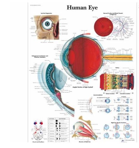
This laminated chart – 20″ wide x 26″ high – shows cross section of the eye.
Chart contents
- Provides lateral and top view of the eye and shows the visual field
- Illustrates anterior chamber angle, lens, retina, fundus and the macula lutea
This educational chart is ideal for patient understanding in a GP surgery or physiotherapy clinic.
Charts are supplied individually.