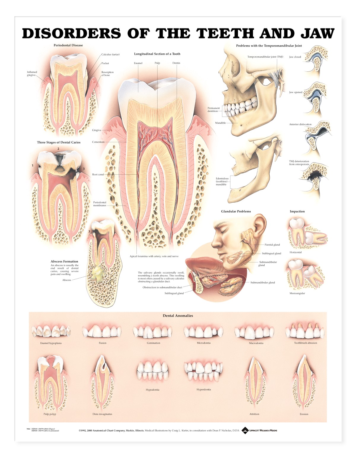
This laminated anatomical chart – 20″ wide x 26″ high – shows longitudinal section of a normal tooth. Illustrates periodontal disease, three stages of dental caries, abscess formation, problems with the temporomandibular joint, glandular problems and impaction.
Chart contents:
- Shows 12 dental anomalies: enamel hypoplasia, fusion, germination, microdontia, macrodontia, toothbrush abrasion, hypodontia, hyperdontia, pulp polyp, dens invaginatus, attrition, and erosion.
This anatomical chart is ideal for patient understanding in a dental surgery.
Charts are supplied individually.